Next tests schedules


Zygotes

Description of 2-PN stage zygotes

The kinetics of PN formation can be followed by time-lapse microphotography. A small movie can be viewed here.

PN centering

The PNs are usually located at or near the center of the zygote but may be peripheral in some zygotes.

EX_PN_POSITION.jpg


PN proximity

After their formation, the female PN moves toward the male PN until they are in close proximity. The 2 PN may appear abutted, in contact or still separated in some zygotes.

EX_PN_PROXIMITY.jpg
PN orientation

The abutted PN rotate in the cytoplasm and their axis may appear aligned with the second polar body or misaligned to a certain extent.

EX_PN_ORIENTATION.jpg
Nucleolar precursor bodies (NPB)

NPB number

The total NPB number may vary from more than 14 to less than 6. A progressive coalescence of NPB is probably responsible for a decrease in NPB number combined with an increase in their size.

EX_NPB_NB.jpg


NPB number asymmetry

Asymmetry in NPB number between the 2 PN is observed in most of the zygotes and probably reflect different patterns of development between the male and female PN. This asymmetry may be however limited or very important.

EX_NPB_SYM.jpg


NPB polarisation

NPB tend to polarise towards the line separating the 2 PN. This polarisation may be very strong, intermediate or totally absent. It may progress differently in the two pronuclei.

EX_NPB_POL.jpg


Cytoplasmic halo

Cytoplasmic organelles, probably mitochondria, move towards the center of the zygote around the 2PN, resulting in a clear halo at the zygote periphery. The size of the halo varies according to the zygotes. It may be absent in some zygotes.

EX_HALO.jpg


Parameters of the EQC test

The EQConline test will give you several possible answers for the various characteristics of a 2 pronuclear zygote. You can make only one choice.
Although you may not be used to grade the zygotes in your day-to-day activity, grading is a convenient way of communicating zygote characteristics both to colleagues and medical staff. In our view, it this is an important parameter to monitor in an External Quality Control scheme.


PN position

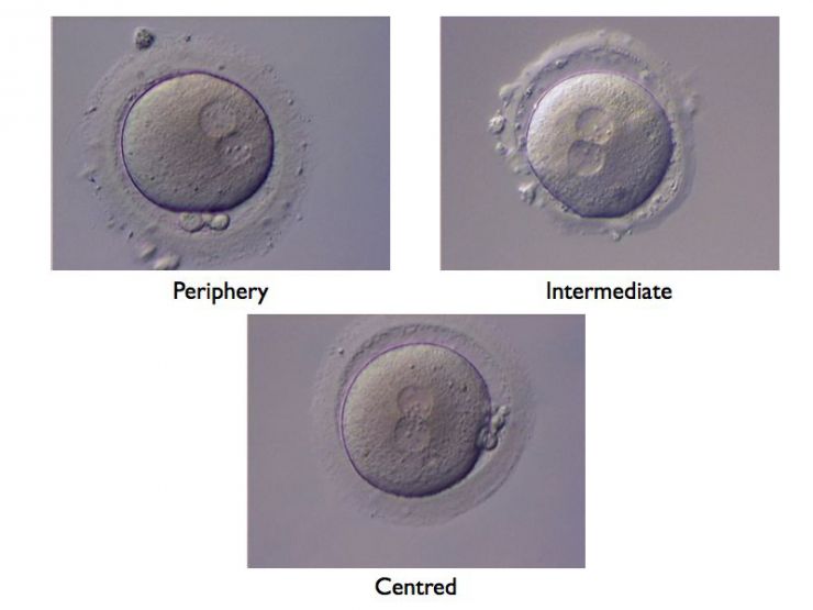
The position of the pronuclei (PN) change over time. They appear usually at the periphery where the spermatozoon entered into the oocyte. The aster created by the sperm centriole will bring the female and male pronuclei together and the two will the migrate together towards the center of the oocyte, where the events leading to the first mitotic division will take place.


Parameters Morphological aspects
Periphery clearly situated near the ooplasm
Intermediate no more in the periphery, but still not at the center
Centered in the middle of the cell


PN proximity

The male pronucleus originates from the sperm head, whereas the female pronucleus appears after the second meiotic division. They contain the male and female chromosomes respectively. These two organelles are brought together by the aster originating from the sperm centriole. The two pronuclei may thus appear separated, close or abutted depending at which moment the zygote is observed. One should keep in mind that this is a dynamic process, which does not represent a stable characteristic of the zygote.


Parameters Morphological aspect
Separated the two PN are separated by a some cytoplasmic material
Close the two PN are touching, but have not built yet a common frontier. Depending on the orientation of the oocyte, this stage may be mistakingly taken for the two other ones
Abutted

the two PN have created a common frontier and have lost their spherical appearance


PN sizes

The male pronucleus (the one situated most distantly from the polar bodies) may appear slightly bigger. In most situation, this difference is barely visible and the two pronuclei exhibit sizes that are similar.


Parameters Morphological aspect
Strongly unequal the two PN exhibit sizes that are clearly different
Slightly unequal the sizes of the two PN is slightly different
Equal the two PN are of comparable size


 NPB number

The nucleolar precursor bodies (NPB) are intranuclear structures that will lead to the formation of nucleoles. Their number and sizes vary from species to species, and in humans from zygote to zygote. They are correlated with early RNA synthesis activity. Three classes have been considered here (<6, 6-14, >14).


Parameters Morphological aspect
<6 1-5 NPB counted in the two PN
6-14 6-14 NPB counted in the two PN
>14 more than 14 NPB counted in the two PN

NPB symmetry

The number of nucleolar precursor bodies (NPB) may not be identical in the two PN. Three situations are considered here, where the number of NPB may be equal, different by less or more than 3 units.


Parameters Morphological aspect
∂≤3 the NBP numbers in the two PN differ by 3 or less
∂>3 the NBP numbers in the two PN differ by more than 3
Symmetrical the same number of NPB is found in the two PN


NPB polarisation

The nucleolar precursor bodies may be evenly distributed throughout the PN or may tend to concentrate along the common border between the two PNs. Three situations are considered here.


Parameters Morphological aspect
Non polarised the NPBs are evenly distributed
Polarised the NPBs are aligned along the common border
Intermediate the NBPs are not evenly distributed, but clearly tend evenly or unevenly to concentrate near the common border


Cytoplasmic halo

Following fertilization, the cytoplasm may retract as a result of the migration of the organelles towards the two PN. This leads to the formation of a clear zone at the periphery of the zygote, known as the cytoplasmic halo. The extent of this halo can be large, small or inexistent.


Parameters Morphological aspect
None no cytoplasmic halo is visible
Small a small cytoplasmic halo is visible near the oolemma
Large a large and clearly visible cytoplasmic halo appears on more than one-fourth of the circumference

Effects of zygote phenotypes on embryo development and implantation


Parameters Effect on implantation
PN centring Detrimental if peripheral
PN proximity Detrimental if distant
PN orientation Controversial
NPB polarisation Beneficial if strong
NPB asymmetry Detrimental if important (NPB>3)
Cytoplasmic halo Beneficial

References

  • Ebner T et al. Selection based on morphological assessment of oocytes and embryos at different stages of preimplantation development: a review. Human Reprod Update 9: 251-262, 2003.
  • Garello et al. Pronuclear orientation, polar body placement, and embryo quality after intracytoplasmic sperm injection and in-vitro fertilization: further evidence for polarity in human oocytes? Human Reprod 14: 2588-2595, 1999.
  • Senn A et al. Morphological scoring of human pronuclear zygotes for prediction of pregnancy outcome. Human Reprod 21: 234-239, 2006.
  • Scott L. Pronuclear scoring as a predictor of embryo development. RBMonline 6: 201-214, 2003.
  • Tesarik J and Greco E. The probability of abnormal preimplantation development can be predicted by a single static observation on pronuclear stage morphology. Human Reprod 14: 1318-1323, 1999.